As the “Yellow Eclipse” cast its shadow over my body, I felt the darkness closing in. The jaundice had taken hold, its grip tightening with each passing day. But I refused to surrender. I sought out the warriors of the medical world, armed with their scalpels and their science. They launched a counterattack, unleashing a barrage of treatments that pierced the shadows and illuminated a path forward.
With each passing day, the light grew stronger. The bile ducts, once blocked, began to flow like rivers unchained. The yellow hue that had shrouded my skin started to fade, replaced by a warm, golden glow. I felt the eclipse receding, its power waning as my body began to heal. The treatments were working, and I was rising from the ashes like a phoenix reborn. Hope flickered to life, a flame that burned brighter with each step forward.
Now, I stand bathed in the warm light of triumph, the “Yellow Eclipse” a distant memory. My body, once a battleground, is now a testament to the power of medical miracles. I savor each breath, each beat of my heart, each moment of freedom from the shadows. The journey was long, but the destination was worth it – a life untethered, unbridled, and full of flavor. The taste of victory is sweet, and I am hungry for whatever comes next. Bring on the feast of life!
What is Obstructive Jaundice?

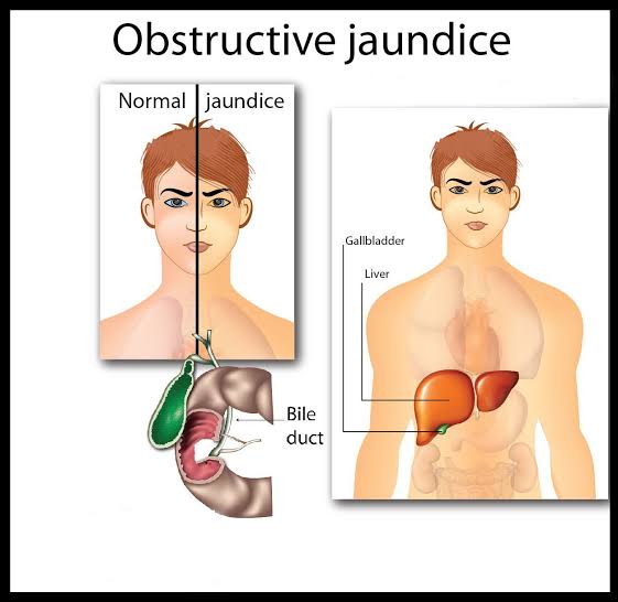
Obstructive jaundiceis a medical condition characterized by a blockage in the bile ducts, which prevents bile from flowing into the intestine. This blockage causes bile to build up in the liver and blood, leading to a yellowish discoloration of the skin and eyes (jaundice).
Causes
Gallstone-related causes:
1. Choledocholithiasis: Gallstones in the common bile duct.
2. Cholecystolithiasis: Gallstones in the gallbladder that can migrate to the bile ducts.
Tumor-related causes:
1. Cholangiocarcinoma: Cancer of the bile ducts.
2. Pancreatic cancer: Tumors that compress or invade the bile ducts.
3. Ampullary cancer: Tumors at the ampulla of Vater (where the bile and pancreatic ducts meet).
4. Metastatic tumors: Cancer that spreads to the liver or bile ducts from other parts of the body.
Inflammatory causes:
1. Primary sclerosing cholangitis (PSC): Inflammation and scarring of the bile ducts.
2. Secondary sclerosing cholangitis: Inflammation and scarring due to infection, injury, or surgery.
3. Chronic pancreatitis: Inflammation of the pancreas that can affect the bile ducts.
Other causes:
1. Bile duct strictures: Narrowing of the bile ducts due to scarring or injury.
2. Parasitic infections: Infections like liver flukes that can block the bile ducts.
3. Congenital anomalies: Rare birth defects that affect the bile ducts.
4. Trauma: Injury to the bile ducts due to surgery, accident, or other causes.
5. Iatrogenic causes: Complications from medical procedures or treatments.
These causes can lead to the blockage of bile ducts, resulting in the symptoms of Obstructive Jaundice.
Types
1. Intrahepatic Obstructive Jaundice: Blockage within the liver, often due to tumors, cysts, or scarring.
2. Extrahepatic Obstructive Jaundice: Blockage outside the liver, commonly caused by:
– Choledocholithiasis (gallstones in the common bile duct)
– Cholangiocarcinoma (bile duct cancer)
– Pancreatic cancer
– Ampullary cancer (tumors at the ampulla of Vater)
3. Posthepatic Obstructive Jaundice: Blockage after the bile leaves the liver, often due to:
– Gallstones
– Tumors (e.g., pancreatic, ampullary, or cholangiocarcinoma)
– Strictures (narrowing of the bile ducts)
4. Benign Obstructive Jaundice: Non-cancerous blockages, such as:
– Gallstones
– Inflammatory strictures
– Congenital anomalies
5. Malignant Obstructive Jaundice: Cancerous blockages, including:
– Cholangiocarcinoma
– Pancreatic cancer
– Ampullary cancer
– Metastatic tumors
Understanding the type of Obstructive Jaundice helps determine the appropriate treatment approach.
Pathophysiology
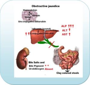
1. Blockage of bile ducts: A obstruction in the bile ducts, either intrahepatic (within the liver) or extrahepatic (outside the liver), prevents bile from flowing into the intestine.
2. Bile accumulation: Bile salts and bilirubin build up in the liver and blood, causing an increase in serum bilirubin levels.
3. Jaundice: The excess bilirubin in the blood binds to albumin and is transported to peripheral tissues, causing yellow discoloration of the skin and eyes (jaundice).
4. Bile salt accumulation: Bile salts, normally excreted into the intestine, accumulate in the blood and tissues, leading to:
– Pruritus (itching): Bile salts stimulate nerve endings, causing itching.
– Fat malabsorption: Bile salts are essential for fat digestion; their absence leads to malabsorption.
5. Liver damage: Prolonged obstruction leads to liver inflammation, fibrosis, and potentially cirrhosis.
6. Increased pressure: Blockage causes increased pressure in the bile ducts, leading to:
– Dilation: Expansion of the bile ducts.
– Rupture: Potential rupture of the bile ducts, causing peritonitis.
7. Infection: Stagnant bile becomes a breeding ground for bacteria, leading to:
– Cholangitis: Infection of the bile ducts.
– Sepsis: Systemic infection.
8. Malnutrition: Malabsorption of fats and fat-soluble vitamins (A, D, E, and K) leads to nutritional deficiencies.
Signs and Symptoms
Common Symptoms:
1. Jaundice: Yellow discoloration of the skin and eyes
2. Dark urine: Tea-colored or amber-colored urine
3. Pale stools: Clay-colored or pale stools
4. Itching (pruritus): Due to bile salt accumulation
5. Fatigue: Feeling weak or tired
6. Weight loss: Unintentional weight loss
7. Abdominal pain: Pain in the upper right or middle abdomen
8. Nausea and vomiting: Feeling queasy or vomiting
Less Common Symptoms:
1. Fever: Elevated body temperature
2. Chills: Feeling cold
3. Loss of appetite: Decreased interest in food
4. Diarrhea: Loose or watery stools
5. Abdominal swelling: Ascites (fluid accumulation in the abdomen)
6. Bruising: Easy bruising or bleeding
7. Confusion: In severe cases, jaundice can lead to encephalopathy (brain dysfunction)
Physical Signs:
1. Yellow skin and eyes
2. Enlarged liver (hepatomegaly)
3. Enlarged spleen(splenomegaly)
4. Tenderness: Abdominal tenderness, especially in the upper right quadrant
5. Ascites: Fluid accumulation in the abdomen
Laboratory Findings:
1. Elevated bilirubin: Increased serum bilirubin levels
2. Elevated liver enzymes: Increased levels of AST, ALT, and alkaline phosphatase
3. Abnormal bile ducts: Ultrasound or imaging studies may show dilated or blocked bile ducts
Complications
Comlications from Blockage:
1. Cholangitis: Infection of the bile ducts
2. Sepsis: Systemic infection
3. Liver abscess: Pus-filled cavity in the liver
4. Hepatic failure: Liver dysfunction or failure
5. Bile duct perforation: Hole in the bile duct, leading to peritonitis
Comlications from Related Conditions:
1. Gallbladder cancer: Increased risk with chronic gallstones
2. Pancreatic cancer: Increased risk with chronic pancreatitis
3. Liver cancer: Increased risk with chronic liver disease
4. Malnutrition: Malabsorption of fats and fat-soluble vitamins
Comlications from Treatment:
1. Surgical complications: Bleeding, infection, or organ damage during surgery
2. ERCP complications: Pancreatitis, bleeding, or perforation during ERCP
3. Stent occlusion: Blockage of the stent, requiring repeat procedures
4. Liver transplantation complications: Rejection, infection, or organ failure
Long-term Complications:
1. Chronic liver disease: Cirrhosis, fibrosis, or liver scarring
2. Bile duct stricture: Narrowing of the bile ducts, requiring repeat interventions
3. Recurrent jaundice: Repeat episodes of jaundice due to ongoing blockage or disease
Prompt diagnosis, effective treatment, and close monitoring can help prevent or manage these complications.
Prevention
Risk Factor Management:
1. Maintain a healthy weight: Reduce the risk of gallstones and liver disease.
2. Eat a balanced diet: Focus on fruits, vegetables, whole grains, and lean proteins.
3. Limit alcohol consumption: Excessive alcohol consumption can lead to liver damage.
4. Avoid smoking: Smoking increases the risk of liver cancer and other diseases.
5. Manage chronic conditions: Effectively manage diabetes, high blood pressure, and high cholesterol.
Proactive Steps:
1. Get vaccinated: Receive hepatitis A and B vaccinations to prevent liver infections.
2. Practice safe sex: Reduce the risk of hepatitis B and other sexually transmitted infections.
3. Avoid sharing needles: Sharing needles increases the risk of hepatitis B and C.
4. Limit exposure to toxins: Avoid exposure to chemicals, pesticides, and heavy metals.
5. Stay hydrated: Drink plenty of water to help flush out toxins.
6. Exercise regularly: Regular physical activity promotes overall health and well-being.
7. Get regular check-ups: Monitor liver function and bile duct health with regular medical check-ups.
Specific Prevention Measures:
1. Gallstone prevention: Maintain a healthy weight, eat a balanced diet, and limit alcohol consumption.
2. Liver cancer prevention: Get vaccinated against hepatitis B, limit alcohol consumption, and avoid smoking.
3. Bile duct injury prevention: Avoid traumatic injuries, and take precautions during medical procedures.
Treatment and Management
Medical Treatment:
1. Fluid management: Intravenous fluids to prevent dehydration
2. Antibiotics: To treat or prevent infections
3. Pain management: Medication to control pain and discomfort
4. Vitamin supplements: Fat-soluble vitamins (A, D, E, and K) to manage deficiencies
5. Ursodiol: Medication to dissolve gallstones or reduce bile salt production
Surgical Treatment:
1. Endoscopic retrograde cholangiopancreatography (ERCP):
– Remove gallstones or tumors
– Insert stents to open blocked bile ducts
2. Surgical exploration: Open surgery to relieve blockages or remove tumors
3. Bile duct reconstruction: Surgery to repair or rebuild damaged bile ducts
4. Liver transplantation: In severe cases, liver transplantation may be necessary
Minimally Invasive Procedures:
1. Percutaneous transhepatic cholangiography (PTC):
– Insert a catheter through the skin to drain bile
2. Percutaneous transhepatic biliary drainage (PTBD):
– Insert a stent to open blocked bile ducts
Lifestyle Modifications:
1. Dietary changes: Fat-free or low-fat diet to manage malabsorption
2. Weight management: Maintain a healthy weight to reduce gallstone risk
3. Avoid alcohol: Limit or avoid alcohol consumption to prevent liver damage
Follow-up Care:
1. Regular monitoring: Periodic blood tests and imaging studies to monitor liver function and bile duct health
2. Endoscopic surveillance: Regular ERCP procedures to monitor for recurrent blockages

